Excavata: Parabasalia, Trichomonadida
реклама
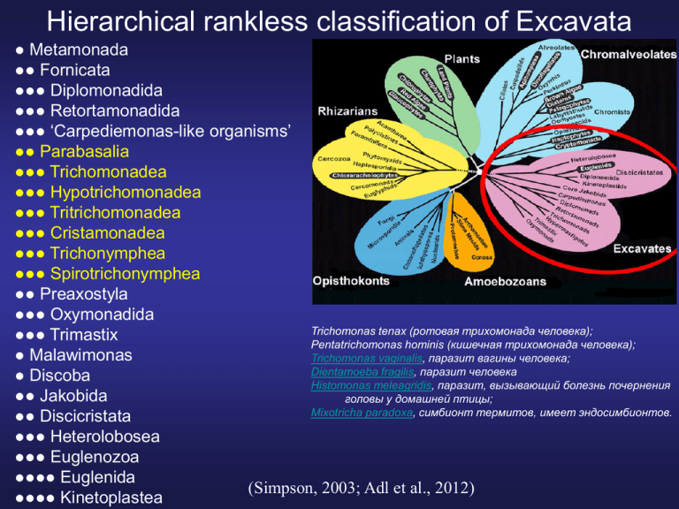
Hierarchical rankless classification of Excavata ● Metamonada ●● Fornicata ●●● Diplomonadida ●●● Retortamonadida ●●● ‘Carpediemonas-like organisms’ ●● Parabasalia ●●● Trichomonadea ●●● Hypotrichomonadea ●●● Tritrichomonadea ●●● Cristamonadea ●●● Trichonymphea ●●● Spirotrichonymphea ●● Preaxostyla ●●● Oxymonadida Trichomonas tenax (ротовая трихомонада человека); ●●● Trimastix Pentatrichomonas hominis (кишечная трихомонада человека); ● Malawimonas Trichomonas vaginalis, паразит вагины человека; Dientamoeba fragilis, паразит человека ● Discoba Histomonas meleagridis, паразит, вызывающий болезнь почернения ●● Jakobida головы у домашней птицы; Mixotricha paradoxa, симбионт термитов, имеет эндосимбионтов. ●● Discicristata ●●● Heterolobosea ●●● Euglenozoa ●●●● Euglenida (Simpson, 2003; Adl et al., 2012) ●●●● Kinetoplastea Proposed evolutionary relationships of parabasalids (Noda et al., 2012). The tree shows the relationships of the six parabasalian classes. Flagellar multiplication in a single mastigont system has occurred independently in the boxed classes. The multiplications have occurred ancestrally in two classes (marked with filled circles) and probably twice within the other class (open circle). Triangles indicate the occurrence of cytoskeletal simplification in undulating membrane (UM) and costa (for details see Noda et al., 2012). The costa has a similar banding pattern in members of Tritrichomonadea and Hypotrichomonadea (A-type striation according to Honigberg et al., 1971), whereas the costa of Trichomonadea shows a different banding pattern (B-type striation). Пельта-аксостилярный комплекс • Пельта – органелла серповидной формы, поддерживающая кинетосомальный комплекс. Из микротрубочек. • Аксостиль контактирует с пельтой – образуется ложкообразное расширение, где лежит ядро. Идет по всей длине тела в виде трубки, заполненной гликогеном (НЗ клетки). Состоит из микротрубочек. Задний конец выдается наружу в виде стержня покрытого плазмалеммой. Аксостиль подвижный. • На границе выхода аксостиля расположено параксостилярное кольцо. Два типа ундулирующей мембраны Lamellar undulating membrane (e.g. Trichomonas, Trichomitopsis, Trichomitus) Специальные подвижные контакты с помощью плотного гомогенного вещества Rail-like undulating membrane (e.g. Tritrichomonas, Monocercomonas) аксонема продольные тяжи параксиальный тяж Ундулирующая мембрана. Парабазальные тела. Кристаллическое тело, представляет собой опорную структуру ундулирующей мембраны. Состоит из множества электронно-плотных, тонких филаментов, расположенных относительно друг друга на равном расстоянии и перпендикулярно. Характерная особенность – парабазальные тела. Состоят из парабазальных фибрил и толстого слоя цистерн аппарата Гольджи (20-30 цистерн в каждом). У многих видов лежат вокруг аксостиля. Митохондрии отсутствуют, но: • Гидрогеносомы –округлые микротельца с плотной мембраной и мелкозернистым матриксом с уплотненной центральной частью. Способны делиться с образованием 2х дочерних органелл. • Две мембраны, но нет внутренних крист. • Одна группа находится возле вогнутой поверхности косты. • Вторая – возле аксостиля. • Тесно связаны с ЭПР и другими органеллами клетки при помощи транспортных пузырьков. • Нет генома. • Нет цикла лимонной кислоты. Продуцируют молекулярный водород в качестве конечного продукта ферментативного расщепления углеводов. • Имеются у некоторых амитохондриатных экскават (Excavata), а именно у парабазалий и у гетеролобазей. Анаэробный метаболизм. to citric acid cycle 1 mol of glucose produces 36 mol of ATP The utilization of the reducing power of ferredoxin is different in the different amitochondriate groups 1 mol of glucose produces 4 mol of ATP Крайне модифицированные митохондрии? Несколько генов, для которых известно митохондриальное происхождение, локализованы в ядре ряда амитохондриальных экскават (HSP10, HSP70, cpn60). Продукты этих генов используются в гидрогеносомах. Размножение • Бесполое размножение. Митоз и цитокинез. Митоз без цитокинеза, образуются многоядерные временные сомателлы (вызывается лекарствами при лечении!). • Закрытый плевромитоз: 1) ядерная оболочка сохраняется, 2) формируется внеядерное веретено. • Пластинчатые атрактофоры и атрактосферидии (большие стрелки) связаны с кинетосомами (перерезанные кинетосомы видны). От атрактофоров отходят микротрубочки веретена и хромосомные микротрубочки. • Хромосомы крепятся к кинетохорам (головка стрелки) в ядерной оболочке. Разнообразие, филогенетические связи Synderella: умножение числа кариомастигонтов Тритрихоманада с ундулирующей мембраной Tritrichomonas Обычная кристоманада Devescovina Joenia: Умножение числа базальных тел Proposed evolutionary relationships of parabasalids (Noda et al., 2012). The tree shows the relationships of the six parabasalian classes. Flagellar multiplication in a single mastigont system has occurred independently in the boxed classes. The multiplications have occurred ancestrally in two classes (marked with filled circles) and probably twice within the other class (open circle). Triangles indicate the occurrence of cytoskeletal simplification in undulating membrane (UM) and costa (for details see Noda et al., 2012). The costa has a similar banding pattern in members of Tritrichomonadea and Hypotrichomonadea (A-type striation according to Honigberg et al., 1971), whereas the costa of Trichomonadea shows a different banding pattern (B-type striation). Parabasalia: Trichomonadida • Исходно 4 жгутика: 3 направлены вперед, 1- назад и может образовывать ундулирующую мембрану. Число жгутиков может быть 4-6. • Кинетосомы 3х жгутиков расположены параллельно друг другу и продольной оси тела, 1 кинетосома направлена под углом. • Корешковая система: 3 коротких фибриллярных корешка, 2 поперечно исчерченные парабазальные фибриллы, коста. • Коста - поперечно исчерченная фибрилла, идет параллельно рекуррентному жгутику, лежит внутри клетки. • Пельта и аксостиль. • У Histomonas только один жгутик и редуцированный аксостиль, а у Dientamoeba и жгутик, и аксостиль отсутствуют. • Яркие представители: Trichomonas vaginalis, паразит половой системы человека. Dientamoeba fragilis, амебоидный паразит человека. Histomonas meleagridis, паразит, вызывающий заболевание «черной головы» у домашних птиц. Mixotricha paradoxa, симбионт термитов, имеет эндосимбионтов. Trichomonadida Thin section showing the recurrent flagellum and the undulating membrane. Bar 0.1 μm Thin section showing the general aspect of the trichomonad. Orientation and location of the nucleus (N), nucleolus (Nu), axostyle (Ax), hydrogenosomes (H), food vacuoles (V), and the recurrent flagellum (RF) with the undulating membrane (UM) can be seen. Dividing hydrogenosomes (DH) can also be observed. Dark dot-like structures are seen following the length of the axostyle. Bar 1 μm. (Rivera et al., 2008) Жизненный цикл трихомонадид SEM of Tritrichomonas foetus during morphogenesis from interphase (Fig. 8 through pre-mitosis (Fig. 9) and mitosis (10–13). Fig. 8. The cell is in interphase, presenting one axostyle tip (arrow) at the posterior region and a teardrop shape. Fig. 9. The cell is larger and shows the flagella already duplicated. Fig. 10. Prophase (or Phase 1). The cell assumes a triangle shape. Fig. 11. The cell is in metaphase, presenting an heart shape. Note the two axostyles tips (white arrows). Fig. 12. Phase 3. The cell is in anaphase. Note the more elongated shape and the appearance of the axostyle tips at the posterior region of the cell (white arrow). The axostyles have started to migrate and in anaphase these tips are apart from each other (arrows, Fig. 12). Fig. 13. Phase 4. The daughter cells are pointing in opposite directions but still connected. The recurrent flagella are in opposite positions (arrowheads). Notice the redirection of the recurrent flagella from a parallel position with relation to the cell body longitudinal axis on premitosis and prophase (Fig. 9–10) to a perpendicular position in relation to the longitudinal axis in Phase 2 (Fig. 11). The axostyle tips are indicated in all phases by white arrows. Asterisk, flagellar channel; F, anterior flagella; R, recurrent flagellum. Figs. 8 and 13, Bar = 3 mkm. Figs 9–12, Bar = 2 mkm. (Ribeiro et al, 2000) Details of the anterior region of Tritrichomonas foetus by SEM, from early prophase 14 (14), to late prophase (16) of mitosis. Notice that in Fig. 14, the flagellar channels (stars) are side-by-side, and gradually they undergo contortion (Fig. 15) and separation (Fig. 16). Figure 14, Bar 5 1 mm. Figs. 15–16, Bar 5 0.5 mm. 17. Computer animation frames of a hypothetical 3D model of the trichomonad’s mitosis. It shows how the cytoskeletal structures combine to promote a karyokinesis coupled to cytokinesis. Prophase (A); metaphase (B); anaphase (C, D) and telophase (E). Spindle microtubules (small arrow), axostyle-pelta complex (a), nucleus (n) costa, which also indicates the recurrent flagella position (c). The anterior portion of Tritrichomonas foetus in longitudinal section by TEM is shown during Phase 2 of mitosis, which corresponds to Fig. 11. Axostyles (arrowheads) are bent due to separation of the uppermost part and proximity of the posterior trunk. The nucleus (N) is positioned in between both axostyles. The recurrent flagella (R) and the costa (C) are seen in crosssection due to their position perpendicular to longitudinal axis of the cell body. Golgi complex (G) and hydrogenosomes (H) are also seen. Bar = 1mkm. (Ribeiro et al, 2000) Hydrogenosome behavior during the cell cycle in Tritrichomonas foetus It is shown that the hydrogenosomes divide at any phase of the cell cycle and that the organellar division is not synchronized. During the interphase the hydrogenosomes are distributed mainly along the axostyle and costa, and at the beginning of mitosis migrate to around the nucleus. Three forms of hydrogenosome division were seen: (1) segmentation, where elongated hydrogenosomes are further separated by external membranous profiles; (2) partition, where rounded hydrogenosomes, in a bulky form, are further separated by a membranous internal septum and, (3) a new dividing form: heart-shaped hydrogenosomes, which gradually present a membrane invagination leading to the organelle division. The hydrogenosomes divide at any phase of the cell cycle. A necklace of intramembranous particles delimiting the outer hydrogenosomal membrane in the region of organelle divisionwas observed by freeze-etching. There are similarities between hydrogenosomes and mitochondria behavior during the cell cycle. Schematic diagram of Tritrichomonas foetus showing the main cell structures. AF, anterior flagella; Ax, axostyle; B, basal body; C, costa; Co, comb; ER, endoplasmic reticulum; G, Golgi; H, hydrogenosomes; I-infra-basal body (infra-kinetosomal body); L, lysosome; N, nucleus; Nu, nucleolus; P, pelta; PF, parabasal filament; R, recurrent flagellum; S, sigmoid filaments; UM, undulating membrane; V, vacuole. (Benchimol, Engelke, 2003) T. foetus in different phases of the cell cycle, showing the hydrogenosomes behavior. 2a. General view of an interphase T. foetus showing the overall organelles distribution. The nucleus (N) is oval and located at the anterior region of the cell. The hydrogenosomes (H) follow the axostyle (A). F, flagella; C, costa; G, Golgi; P, pelta. Bar = 1 μm. 2b. T. foetus in a pre-mitotic phase. The pelta (P) and the axostyle (A) have already duplicated whereas the flagella (F) have not. The hydrogenosomes (H) are seen following both axostyles. Clumps of condensed chromatin are observed in the nucleus (N). Bar = 1 μm. 2c. T. foetus presenting the costa (C) and the recurrent flagellum (RF) already duplicated. The nucleus (N) is elongated, which is characteristic of the mitosis onset. Note that the hydrogenosomes (H) are distributed around the nucleus. A, axostyle. Bar = 1 μm. 2d. T. foetus in late prophase. The cell is triangle shaped and present all cytoskeletal structures already duplicated, such as the costa (C), axostyle (A), and pelta (P). The nucleus (N) shows chromatin clumps and the nucleolus (NU) is present. Note the hydrogenosomes (H) distribution around the nucleus. BB, basal bodies; F, flagella; Bar = 1 μm. 2e. T. foetus in metaphase. The two axostyles (A) are in mirror symmetry, the two Golgi (G) are present and do not fragment, and the elongated nucleus (N) is under constriction. The hydrogenosomes (H) are aligned along the axostyles. C, costa; F, flagella. Bar = 1 μm. 2f. Karyokinesis is just finished and the two nuclei (N) are seen already separated. The chromatin clumps are still evident, and the axostyles (A) are seen in mirror image symmetry. Note that several hydrogenosomes (H) follow the axostyle, although some of them are dispersed (arrowheads). Bar = 1 μm. (Benchimol, Engelke, 2003) Trichomonas, прикрепленный к эпителиальным клеткам вагины. Прикрепленные формы принимают амебоидную форму. Scanning electron micrograph of Trichomonas vaginalis parasites (gray-green) adhering to vaginal epithelial cells (pink). Attached parasites are flattened and amoeba-like; parasites that do not adhere are pear-shaped. (Science, cover of vol. 315, issue 5809 Antonio Pereira-Neves and Marlene Benchimol Santa Ursula University, Rio de Janeiro, Brazil) Phagocytosis To analyse the phagocytic ability and capacity, two isolates of T. vaginalis presenting different virulence grades were used. Complementary techniques, such as fluorescence microscopy, computerbased fluorescence analysis, scanning and transmission electronmicroscopy and the use of drugs that interfere with the actin microfilaments, were used in order to follow the behaviour of the actin cytoskeleton during phagocytosis of yeast cells by T. vaginalis. It was concluded that: (1) T. vaginalis changes its shape rapidly and engulfs the yeast cells, which are almost as large as the parasite; (2) long-term and fresh cultures are able to phagocytose, although the low-virulence strain JT demonstrated a lower activity when compared with the highly virulent T016 isolate; (3) the T016 strain exhibited an amoeboid morphology during the internalization of yeast cells in contrast with the JT strain; (4) attachment of yeast cells to the parasite occurs via the whole cell surface, including both anterior and recurrent flagella; (5) two forms of phagocytosis were observed: a ‘sinking’ process without any apparent participation of plasma membrane extensions and the classical phagocytosis where pseudopodia are extended toward the target cell; (6) the internalized S. cerevisiae are digested in lysosomes; (7) competitor sugars D-mannose or L-fucose inhibit the phagocytosis, and inhibition was 1.67 times higher in long-term cultured JT than that of the parasites from fresh isolate T016; (8) a thick layer of actin microfilaments was present underlying the plasma membrane, and especially in the pseudopodia and around the phagocytosed particles; (9) a dramatic change in the distribution pattern of fibrillar actin occurred during phagocytosis; (10) cytochalasin D depressed the phagocytosis; (11) a non-specific recognition and phagocytosis of yeast cells by T. vaginalis is mediated by a mannose receptor present on the parasite surface; (12) the phagocytic process may occur simultaneously during mitosis of the parasite. (Pereira-Neves, Benchimol, 2007) SEM showing the attachment of S. cerevisiae (Y) to the anterior (a–c) and recurrent flagella (d–f) of fresh T. vaginalis isolate (T016 strain) after 1 h of incubation at a parasite/yeast ratio of approx. 1:20. Note that in (c) and (f), a strong cell-to-cell recognition between the yeast cell wall and the flagellar membrane was established. Similar results were found in the JT strain. AF, anterior flagellum; Ax, axostyle; RF recurrent flagellum. Scale bar, 4 μm. Phagocytosis SEM showing two forms of phagocytosis of yeast cells (orange) by the JT strain of T. vaginalis (green) The first form of phagocytosis involves a ‘sinking’ process, without any apparent participation of plasma membrane extensions (a–c). The second form resembles the classical phagocytosis, as described for professional phagocytes, where pseudopodia (asterisks) and filopodia (arrowhead) extend towards the target cell (d–f), which is subsequently engulfed (white arrows) (d). Note that in (b) and (f) two yeast cells are being internalized simultaneously. In (d), it can be observed that the yeast cell is internalized by a dividing parasite. Notice that the dividing parasite has duplicated structures, such as two anterior flagella and two recurrent flagella. These two phagocytic processes were also observed in the T016 strain. The phagocytosis assay was performed at parasite/yeast ratio of 1:20 after 2 h of interaction. AF, anterior lagellum; Ax, axostyle; RF recurrent flagellum. Scale bar, 4 μm. (Pereira-Neves, Benchimol, 2007) Phagocytosis SEM of a T. vaginalis T016 (green) phagocytosing yeast cells (orange) Steps involved in the internalization of the yeast cells are observed. The experiment was performed at parasite/yeast ratio of 1:20 after 2 h of interaction. The parasite is able to phagocytose yeast cells and bacteria (pink) over the entire cell surface by a ‘sinking’ phagocytic process. An increased number of yeast cells attached to parasites and an amoeboid shape of these parasites when compared with those of long term growth JT isolate can be observed. In (a), several yeast cells are in process of simultaneous internalization. In Figure (c), four yeast cells are seen completely internalized (white asterisks). AF, anterior flagellum; RF recurrent flagellum. Scale bar, 4 μm. (Pereira-Neves, Benchimol, 2007) Phagocytosis TEM shows the steps of yeast cell internalization by the long-term cultured JT strain The parasite/yeast ratio was 1:20, and the cells were co-incubated for 1 h. After the initial contact, the yeast cells (Y) are engulfed by parasite pseudopods (arrows) (a). Notice that pseudopods (arrows) extend (b) further until they completely internalize the yeast cells (c). Cytochemistry for acid phosphatase showed positive reaction in phagocytic vacuole (d), thus confirming the degradation of the yeast. The organelle exclusion zone (asterisks), which corresponds to microfilaments network along the pseudopodia (arrows) (a and b), and around the vacuoles containing morphological intact yeast cells (b), is clearly distinguished. In (a) and (c), it is possible to observe that the yeast cells are internalized by dividing parasites; the parasites contain duplicated structures, such as two nuclei. ER, endoplasmic reticulum; G, Golgi; H, hydrogenosome; L, lysosome; N, nucleus; RF, recurrent flagellum. Scale bar, 500 nm. (Pereira-Neves, Benchimol, 2007) Мочеполовой трихомоноз (трихомониаз) (шифр по МКБ 10 — А.59) • Trichomonas vaginalis. Хорошо изученный вид. 5 жгутиков (4+1) • Очень обычный паразит человека, до 3.5% людей в мире. • В основном, мягкое течение заболевания, но может быть опасным при раке или иммунодефиците (ВИЧ, СПИД, врожденный иммунодефицит). • Поражает у женщин уретру, вагину и другие отделы половой системы; у мужчин — уретру, предстательную железу и придатки яичек. • Аксостиль может служить для прикрепления к эпителию, вызывает механическое разрушение тканей. • Способны к амебоидной трансформации. • Адгезия на эпителии, лизис клеток хозяина (адгезины, цистеин-протеиназы, гиалуронидаза). • Инкубационный период: 7-10 дней или более. • Формы болезни: острая, хроническая, носительство. • Осложнения. T. vaginalis способна фагоцитировать гонококки, хламидии и другие микроорганизмы, которые сохраняют в них жизнеспособность и находят защиту от действия антибиотиков. Отсюда возможность возникновения рецидивов других болезней. Нарушение репродуктивной функции. • Источник инвазии – человек. Путь передачи – половой, сохраняется возможность через фомиты. • Лечение проводят у всех половых партнеров, даже если у некоторых из них клинические проявления отсутствуют. Прогноз благоприятный. Жизненный цикл Trichomonas vaginalis Кишечный трихомоноз (трихомониаз) (шифр по МКБ10 — А07.8 ) Pentatrichomonas (Trichomonas) hominis. Обычно имеет пять передних жгутиков, один из которых направлен назад. У некоторых особей обнаруживается лишь четыре или три передних жгутика. Кроме того, один жгутик идет вдоль ундулирующей мембраны и выходит за ее пределы как свободный рулевой жгутик. Цист не образует. В толстом отделе кишечника человека. Размножение усиливается при диете, богатой клетчаткой и другими углеводами, а также при различных заболеваниях, сопровождающихся диареей. Питаются бактериями, способны фагоцитировать эритроциты. Относят к условнопатогенным организмам, нередко обнаруживаются в испражнениях здоровых людей. Под действием ряда факторов могут приобретать патогенные свойства и вызывать манифестные формы кишечного трихомоноза, которые протекают в форме колитов и энтероколитов. Осложнения: печеночные абсцессы. Прогноз благоприятный. Механизм передачи: фекальнооральный. Пути передачи: загрязненные пища и вода. Tritrichomonas foetus • Похож на Trichomonas, но ундулирующая мембрана более выражена. • Вызывает бычий тритрихомониаз, очень важное венерическое заболевание скота. • Заболевание может быть фатальным, вызвать выкидыш плода, бесплодие и т.д.у скота. Trichomonadida: Mixotricha paradoxa • Trichomonadida. • Has 4 flagella, but looks as if it had many more. • Covered with symbiotic epibacteria, mostly spirochetes. Dientamoeba fragilis Parabasalia, Trichomonadea, Trichomonadidae, Tritrihomonadinae Monocercomonadidae D. fragilis growing in Robinson’s culture, showing ingested rice starch and fine leaflike pseudopodia. Magnification, 400. Iron-hematoxylin-stained smear of D.fragilis showing pleomorphic trophozoites. Note the characteristic fragmented nuclei and the very small mononucleated trophozoite in the center; magnification, x1,000. (Johnson et al., 2004) Electron micrograph of a mononucleated trophozoite of D. fragilis. The nucleus (Nu) and chromatin are labeled. Electron-dense microbody-like inclusions (Mb) surround the nucleus. Digestive vacuoles (Dv) can be clearly seen, one of which contains an ingested bacterium (B). Magnification, 5,200. Electron micrograph of a binucleated trophozoite of D. fragilis. Digestive vacuoles (Dv) contain either myelin or rice starch (Rs). Magnification, 25,000. (Johnson et al., 2004) Electron micrograph demonstrating chromatin bodies (Ch) in the nucleus (Nu), surrounded by a double nuclear membrane. Note the microbody-like (Mb) inclusions, digestive vacuole (Dv), ingested bacteria (B), and Golgi apparatus (Go). Magnification, 15,500. Electron micrograph of D. fragilis demonstrating digestive vacuoles (Dv) containing myelin. Magnification, 21,000. (Johnson et al., 2004) Electron micrograph of D. fragilis exhibiting exocytosis. Magnification, 28,500. Electron micrograph of D. fragilis exhibiting phagocytosis. Magnification, 6,600. (Johnson et al., 2004) Dientamoeba fragilis Parabasalia, Trichomonadea, Trichomonadidae, Tritrihomonadinae D. fragilis growing in Robinson’s culture, showing ingested rice starch and fine leaflike pseudopodia. Magnification, 400. Обитает в толстом кишечнике человека, шимпанзе, макак и некоторых других видов обезьян, а также у овец. получены убедительные данные, свидетельствующие об их роли в развитии энтероколитов Прогноз благоприятный. Источник инвазии — человек, другие животные (?). Механизм передачи: ?
